Definition :
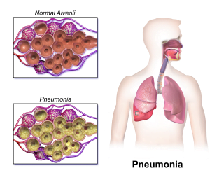
Pneumonia is an infection of the lung parenchyma (ie alveoli rather than bronchi) of infective origin. It is Most common infectious cause of death.
Usually characterized by consolidation:
Consolidation is a pathological process in which ah the alveoli are filled with a mixture of inflammatory exudate, bacteria and WBC.
Classification :
i. Anatomical
- Lobar pneumonia
-
Bronchopneumonia
ii .Based on the basis of acquisition
-
Community – acquired pneumonia (CAP)
-
Aspiration Pneumonia
-
Pneumonia in immunocompromised
Pathophysiology
i. Microbial invasion of the normality- sterile
lower respiratory tract
ii. Three routes:
-
Inhaled aerosolized particles
-
Haemategenous spread
-
Aspiration of oropharyngeal contents
Aspiration of secretions from the airway is the main source of infection for VAP in mechanically-ventilated (MV) patients, and infection develops when bacteria overwhelms the host’s defences.
Lobar pneumonia
Acute bacterial infection of a part of lobe lungs or entire lobe or even two lobes of one or both the lungs
Chest X-ray for lobar pneumonia:
-
Consolidation confined to one or more lobes(or segment of lobes) of lungs
Bronchopneumonia
Infection of the terminal bronchioles that extends into the surrounding alveoli resulting in patchy consolidation of the lungs
- Chest X Ray: Patchy consolidation usually in the base of both lungs.

Community acquired Pneumonia:
Pneumonia which develops in an otherwise healthy person outside of hospital or have been in hospital for less than 48 hour
Risk factors:
-
Comorbidity: nephrotic diseases neurological problem
- Alcoholism
- Advanced age
- Asthma
- Immunosuppression
Etiology:
Potential etiological agents in CAP:
- Bacteria
- Virus
- fungi
- protozoa
Potential bacteriologic cause can be divided into two types:
i. Typical bacterial pathogens
-
Streptococcus pneumonia- 30-60%, severe illness death
-
Haemophilus influenzae – 10%
-
Staph. Aureus (in selected patients)
Gram negative bacilli: klebsiella pneumonia, pseudomonas aeruginosa
ii. Atypical bacterial pathogens
-
Mycoplasma pneumoniae
-
Legionella pneumoniae
-
Viral pneumoniae
-
Fungal pneumoniae
Clinical Features: (Sign & Symptoms)
- Incident to fulminant in presentation
- Mild to fatal in severity
i. Typical symptoms
- Fever with chills and rigors
-
Cough
-
Rust coloured sputum
-
Mucopurulent sputum
-
Dyspnea
-
Pleuritic chest pain
Sign:
- Increased respiratory rate and use of accessory muscles of respiration
- Palpation may reveal increased or decreased tactile fremitus and the percussion note
- Crackles bronchial breath sounds and possibly a pleural friction rub maybe heard on auscultation
- Septic shock, hypotension, multiorgan failure in late stage
Investigation:
- CBC, ESR, CRP
- Sputum analysis
- Chest X Ray- consolidation
- ABG if spo2 is less than 90%
- Thoracocentesis
- Bronchoscopy
- Pleural biopsy
Management:
General:
- Oxygen
- Rehydration
- Treatment of pleural pain
- Pethidine 50 mg iv/im or morphine 10 mg iv/im
- physiotherapy
Specific
-
Antibiotics
-
Beta lactam lantibiotics- penicillin
-
Respiratory fluoroquinolones
-
Macrolides- erythromycin
-
Antipseudomonal beta lactam- Vancomycin
-
Carbapenems
Hospital Acquired Pneumonia (HAP):
Pneumonia that was not incubating upon admission and developing in a patient hospitalized far greater than 48 hours
Predisposing factors:
-
Reduced host defence against bacteria
-
Reduced immune defence (corticosteroid t/t, diabetes, malignancy)
-
Reduced cough reflux (post operative)
-
Reduced mucocilliary clearance (Anaesthetic agents)
-
Aspiration of nasopharyngeal or gastric secretions
-
Immobility or reduced concious level
-
Vomiting, dysphagia
-
Nasogastric intubation
-
Most common pathogen-aerobic gram negative bacilli
- Most common exposed to multiresistant hospital pathogen
- 86% nosocomial infection- Mechanical ventilation
- Mortality – 0 to 50%
Source of infection:
-
Bacterial introduction to lower respiratory tract
-
Endotracheal intubation
-
Infected ventilation/ nebulizer/bronchoscopy
-
Dental or sinus infection
-
Bacteraemia
-
Abdominal sepsis
-
Intravenous canula
Causative Organism :
Common organisms:
Gram negative bacteria
- Escheria coli
- Kliebsiella species
- Pseudomonas aeurginosa
Gram positive bacteria
- Streptococcus pneumoniae
- Staphylococcus pneumoniae
Less common organism
-
Gram negative bacilli: Enterobacter species, Proteus species
-
Fungal : Aspergillus fumigatus
-
Viruses : Cytomegalovirus, herpes simplex
Sign and symptoms of hospital acquired pneumonia:
- Fever with chills and rigors
- Cough
- Rust coloured sputum
- Mucopurulent sputum
- Dyspnoea
- Pleuritic chest pain
Sign:
- Palpation may reveal increased or decreased tactile fremitus and the percussion note.
- Increased respiratory rate and use of accessory muscles of respiration.
- Cyanosis
- CXR- mottled opacities in both fields
Investigations :
-
CBC, ESR, CRP
-
Sputum analysis
-
Chest Xray- consolidation
-
ABG if Spo2 is less than 90%
-
Thoracocentesis
-
Broncoscopy
-
Pleural biopsy
Management of HAP:
Specific
-
Metronidazole 500 mg iv tds plus co-amoxiclav 1.2 gm iv TDS
-
Third generation cephalosporins
-
Meropenem/aztreonam plus flucloxacillin
General
- Oxygen
- Rehydration
- Treatment of pleural pain
- Pethidine 50 mg iv/im or morphine 10 mg iv/im
- physiotherapy
Complications :
i. Pulmonary
-
Collapse
-
Lung abscess
ii. Pleural
-
Pleural effusion
-
Empyema
-
Pneumothorax
iii.Airway
-
Bronchiectasis
-
Sinusitis
Iv. Cardiovascular
- Pericarditis
- myocarditis
- Endocarditis
-
Atrial fibrillation
V. Systemic
- Septicaemia/sepsis
- Hypotension
- Respiratory failure

Your blog is a true hidden gem on the internet. Your thoughtful analysis and engaging writing style set you apart from the crowd. Keep up the excellent work!